Anatomy of Blood
Blood: type of fluid connective tissue that circulates throughout the body, carrying substances to and away from bodily tissues
pH 7.4
Blood is composed of elements…
Red blood cells -
White blood cells
Platelets
Lymph nodes that are enriched in lymphocytes and macrophages that provide surveillance by the immune system.
Leukocytes are white blood cells that help guard against infection.
Large number of leucocytes and lymphocytes are enriched in lymph nodes, where they monitor and respond foreign molecules washed into the system.
Plasma contains nutrients, hormones , antibodies and other immune proteins.
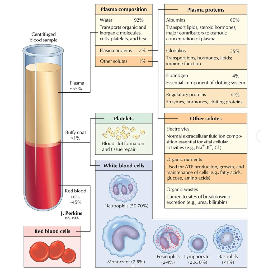
5-7% of carbon dioxide is dissolved in plasma
Plasma carries RBC, WBC, platelet cells throughout the body.
Plasma helps control body temperature and transport substances.
Plasma: About 92% water
55% of whole blood
Buffy coat: consists of white blood cells and platelets, layer found between reddish mass and plasma layers
Leukocytes and platelets
<1% of whole blood
Erythrocytes:
45% of whole blood
Functions of Blood
The main functions of blood is to transportation, regulation, and protection
Blood transports the following throughout the body…
Gases: blood delivers oxygen from the lungs to all cells in the body. Also transports carbon dioxide to the lungs for elimination from the body
Nutrients: blood transports nutrients from the digestive tract and storage sites in the body to various places in the body
Wastes: blood transports waste products to the liver, where they are excreted as bile. Waste products also travel by blood to the kidneys when they need to excreted as urine
Hormones: blood transports hormones from the glands where they are produced to their target organs
Primary function of blood is to distribute substances throughout the body but it also helps regulate the body temperature (maintained with plasma and speed of blood flow), chemical balance, and water balance
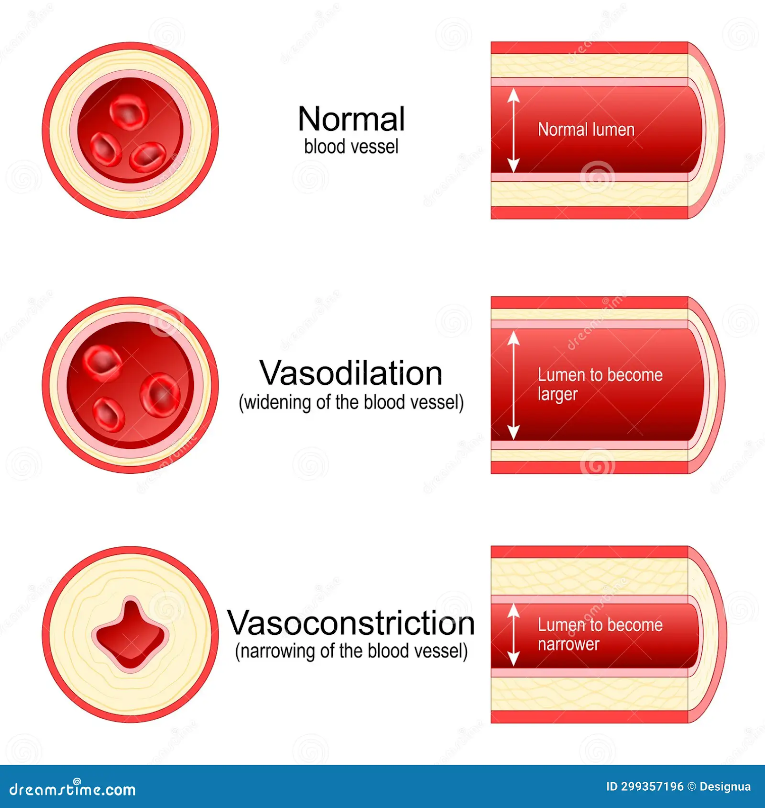
Vasodilate: when blood vessels expand
Occurs when the temperature of the external environment is high
Causes heat loss
Vasoconstrict: blood vessels contract
Occurs when external environmental temperatures are low or blood vessels contract
Less heat is released
Blood helps defend the body against the body against pathogens (foreign invaders)
Blood carries WBC and antibodies to destroy the pathogens
Platelets + Plasma proteins help blood protect body from extensive blood loss if blood vessel is damaged
Hemostasis
Hemostasis: maintain blood in its fluid state and stops blood from leaking out of damaged vessel though clot formation
Steps of Hemostasis:
Vascular spasm/vasoconstriction: blood vessels constrict to reduce blood loss, reduces blood loss for several hours, process works best with small blood vessels
Platelet Plug formation: adhere to the epithelial wall of the blood vessel and aggregate by sticking together creating a temporary seal over damaged site
Blood coagulation: or blood clotting, a series of events that strengthen the platelet plug by using fibrin threads to form mesh around plug, protein mesh= molecular glue, securing the plug to the damaged site
RBC and platelets remain trapped at damaged site, forming a clot that facilitates wound healing
Blood Grouping and Agglutination
Blood types: A, B, AB, and O
Classified based on RBC antigens found on surface of a red blood cell
Types of antibody in blood also identifies a particular blood group, Antibodies: are proteins found in plasma, function as part of the body;s natural defense to recognize foreign substance and alert the immune system
Blood group A: displays type A antigens on the surface of RBC and contains B antibodies on the plasma
Blood group B: Displays type B antigens on RBC surface and contains A antibodies in the plasma
Blood group O: Does not display A or B antigens on the surface of a RBC. Both A and B antibodies are in the plasma
Blood group AB: Displays type A and B antigens on the RBC surface, but neither A nor B antibodies are in the plasma
Rh Factor: protein that exists on RBC surface, protein can be present (+) or absent (-), A+, A-, B+, B-, O+, O-, AB+, and AB-
Agglutination: clumping
Cardiovascular system
Ciculates substances throughout the body using the body using blood as a transporting mechanism
Organs of cardiovascular system work together to supply cells and tissues with oxygen and nutrients and remove cellular wastes such as CO2
* Blood, heart, blood vessels form this system
Blood circulation is closed loop system so blood is contained within the heart or blood vessels at all times
Three types of blood vessels:
Arteries: carry blood away from the heart, toward organ and tissues
Veins: carry blood toward the heart, away from organs and tissues
Capillaries: tiny vessels that form a network around tissues
Arterioles: further divide into capillaries
Veins branch into venules before further diving into capillaries
Heart is located between the lungs in the middles of chest, rests behind and slightly to the left of the sternum or breastbone
Heart is a muscular organ composed of cardiac muscle
There are four chambers:
Two upper chambers atria
Atria are separated from the ventricles by a muscular structure (Septum)
Two lower chambers ventricles
Three layers make up the heart wall:
Pericardium: outer layer
Myocardium: middle layer
Endocardium: innermost layer
Four heart valves that regulate blood flow into and out of the heart:
Tricuspid valve: regulates blood flow between the right atrium and right ventricle
Pulmonary valve: regulates blood flow from the right ventricle into the pulmonary artery
Mitral valve: regulates blood flow from the left atrium into the left ventricle
Aortic valves: regulated blood flow from the left ventricle to the aorta
Aorta: largest artery in the body
Circulation and the Cardiac Cycle
-Blood flows in one direction
Systemic circulation:
Pulmonary vein pushes oxygenated blood into the left atrium
As atrium relaxes, oxygenated blood drains into the left ventricle through the mitral valve
The left ventricle pumps oxygenated blood to the aorta
Blood travels through the arteries and arterioles before reaching the capillaries that surround the tissues
Pulmonary circulation:
Two major veins, superior vena cava + inferior vena cava, brings deoxygenated blood from the upper and lower half of the body
Deoxygenated blood is pooled into the right atrium and then sent out into the right ventricle through the tricuspid valve, which prevents blood from flowing backward
The right ventricle contracts, causing blood to be pushed through the pulmonary valve into the pulmonary artery
Deoxygenated blood becomes oxygenated in the lungs’
Oxygenated blood returned from the lungs into the left atrium through the pulmonary veins
Cardiac cycles: complete cycle beginning with atrial contraction and ending with ventricular contraction
Systole: heart contracts and pumps blood into systemic circulation
Diastole: period of relaxation when the heart chamber fill with blood
EKG: electrocardiogram is a graph that shows the heart’s rate and rhythm over a period of time
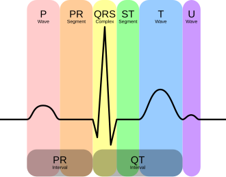
P wave: first wave, indicates serial contraction or systole
QRS wave: indicated ventricular systole and contraction
T wave: indicates ventricular diastole
Flat line between S + T wave is ST segment
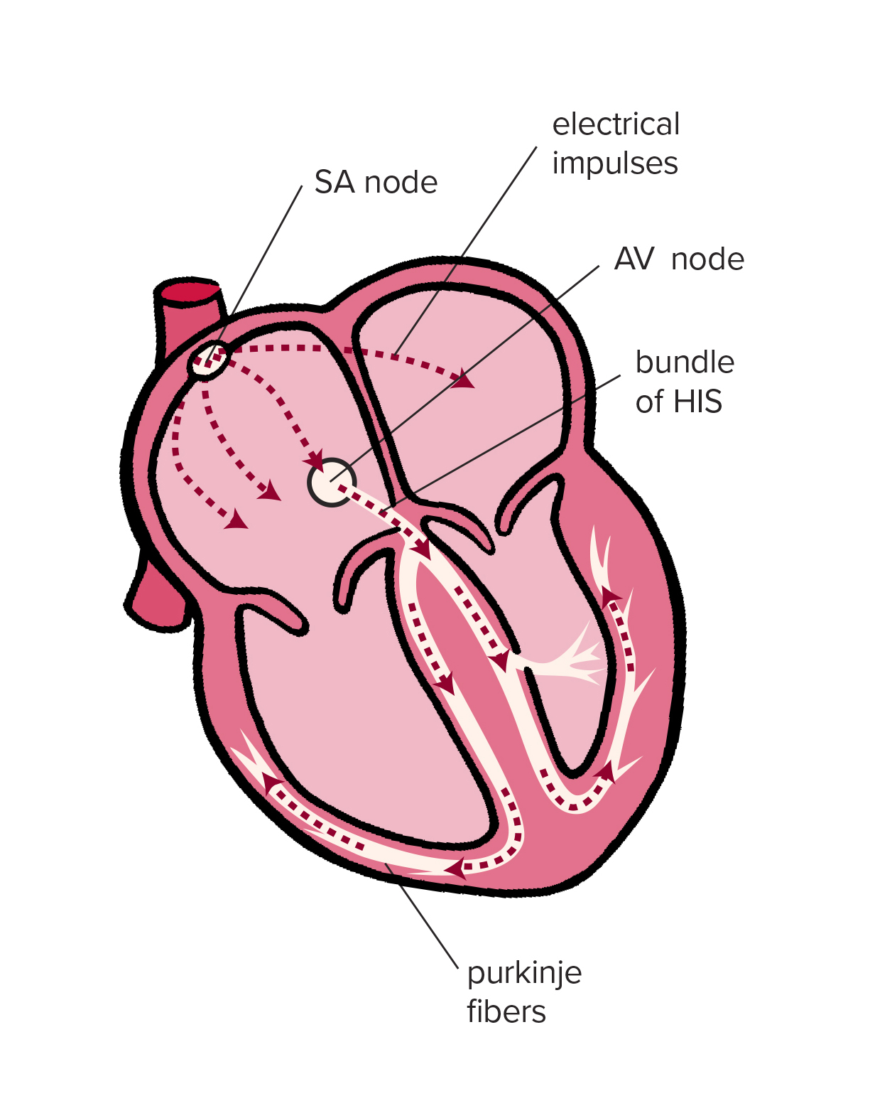
SA node or SInoatrial node
Contraction are controlled by pacemaker called sinoatrial node
Function of cardiovascular system
Perform the vital functions of transporting nutrients, hormones and waste.
Structure of the heart
Heart is made up of cardiac muscles
Split four chamber
Upper chamber is atria and lower chamber is ventricles
There are two large integrated circulatory systems .
The close circulatory system is a double loop system consisting of thick walled arteries that transport blood away from the heart .
Closed double-loop system transports blood ,hormone.
There are two parts of loop: The pulmonary and systemic
Pulmonary carries deoxygenated blood from the right ventricle to the lungs where it is oxygenated and returns oxygenated blood to the left atrium
Right ventricle : The right ventricle pumps blood toward the lungs.
Left ventricle : The left ventricle pumps blood to the body.
Pathology of circulatory system
Heart attack
Stroke
Aneurysms
Atherosclerosis
Arrhythmias
Hypertension
The heart is made up of four chambers, two atria and two ventricles.
Blood cells travel through the heart chambers.
Vein
Veins transport blood from the lungs or the body to the heart.
Veins can carry oxygenated or deoxygenated blood.
No comments:
Post a Comment